Your sinusitis symptoms are persistent. Your doctor mentions testing, but which one: an endoscopy, a CT scan, or both? What’s the difference, and does it really matter?
Both are diagnostic tools, but they serve different purposes, and understanding that difference matters. The answer depends on what your doctor needs to see and how detailed that picture needs to be. By the end of this post, you’ll understand why each test exists and when it’s the right choice for your situation.
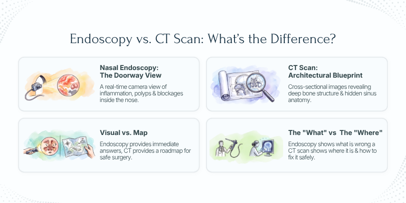
Nasal Endoscopy vs. CT Scan: What's the Difference?
Think of it this way: nasal endoscopy is like looking in the door, while a CT scan is like getting an architectural blueprint.
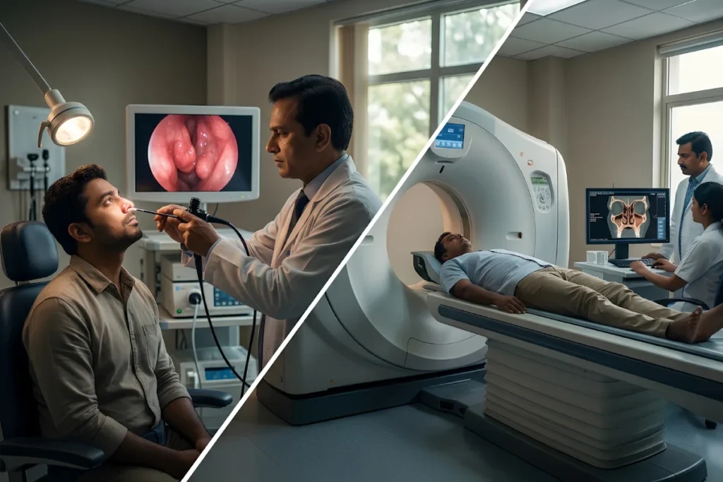
Nasal endoscopy uses a thin, flexible camera that your doctor guides into your nose and sinuses. You get a real-time, detailed view of what’s happening inside: inflammation, blockages, polyps, and drainage patterns visible on a monitor. The findings are immediate. Your doctor explains them while you’re still in the office.
A CT scan works differently. A machine takes cross-sectional images of your sinuses from outside your body. These images reveal bone structure, hidden areas deep in the sinuses, your overall anatomy, and how everything is arranged. You receive the images but wait for a radiologist’s report.
Different information answers different questions. Endoscopy shows what’s wrong. CT shows where it is and how to fix it safely. Both have value, and sometimes you need both.
Nasal Endoscopy: Your First Diagnostic Tool
In most cases, endoscopy is your starting point.
It reveals swelling and inflammation in nasal tissue, shows nasal polyps in detail: their size, location, and severity. It identifies obstruction from a deviated septum or enlarged turbinates. It shows pus, infection, and drainage patterns. Whether inflammation is confined to one sinus or spread throughout becomes clear immediately.
When is it ordered first? During your initial consultation for acute or chronic sinusitis symptoms. If you walk in with congestion, drainage, or breathing difficulty, endoscopy provides immediate answers. There’s no waiting for imaging results or radiology reports. Your doctor explains findings in real-time.
The advantages are significant. You get results instantly, without radiation exposure. It’s cost-effective as a screening tool. And yes, it’s uncomfortable for roughly 30 seconds, but not painful.
Clinically, I start with endoscopy in nearly every patient. It tells me whether surgery is even necessary or if conservative treatment might resolve your symptoms. Consider a patient with heavy nasal congestion. Endoscopy reveals severe turbinate hypertrophy. We immediately know what’s causing the blockage. That clarity directs the next step.
CT Scan: When You Need the Full Picture
CT becomes essential when surgery is being considered or when endoscopy findings don’t fully explain persistent symptoms.
A CT scan reveals your bone structure and sinus anatomy. It shows exactly how severe a deviated septum is and its precise location. It maps sinus dimensions and air-cell patterns. Most importantly, it reveals inflammation deep in the sinuses that endoscopy can’t always visualize. It can rule out rare but serious complications, such as infection spreading beyond the sinuses.
Before sinus surgery, a CT scan is nearly always ordered. It becomes your surgical blueprint. It guides which approach is safest and most effective. For recurrent sinusitis, CT helps explain why symptoms keep returning by revealing anatomical variations that affect treatment. Before revision surgery, a new CT is needed because previous surgery changed your anatomy.
The radiation concern is legitimate. Yes, CT involves radiation exposure, but the diagnostic benefit outweighs this minimal risk. Modern CT protocols minimize exposure while maintaining image quality.
Here’s a real-world example: A patient needs FESS surgery. Endoscopy shows polyps, but the CT scan reveals a narrowing in the ostiomeatal complex that explains why sinusitis keeps recurring. This finding changes our entire surgical strategy.
Endoscopy, CT, or Both? The Decision Logic
The typical pathway is straightforward. Most patients receive endoscopy first during diagnosis. If surgery becomes the recommended path, a CT scan follows for surgical planning. Occasionally, both are done simultaneously in complex or revision cases.
Why both? Endoscopy answers “What’s wrong?” while CT answers “Where is it, and how do I fix it safely?” Together they create a complete diagnostic picture and treatment roadmap.
Cost concerns are understandable. Two tests costs more than one. But a surgeon operating without a CT scan is like a carpenter building without measuring. The right tool prevents complications and wasted time. Typically you’ll have endoscopy during your consultation, then a CT within one to two weeks if surgery is planned.
Sometimes one test is enough. If conservative treatment is your path forward, endoscopy provides sufficient diagnosis. Your doctor’s recommendation reflects your specific case, and it’s fair to ask why each test is being ordered.
How I Approach Testing in Practice
My approach is systematic. Every patient starts with endoscopy. It’s informative, quick, and radiation-free. It tells me what’s happening.
If surgery emerges as the right choice, I order a CT scan. That scan becomes my surgical roadmap. It shows exactly where inflammation lives, how your anatomy is arranged, and what approach is safest and most effective.
Some patients ask whether we can skip the endoscopy and just do the CT. Technically possible, but you’d pay for imaging you don’t need if conservative treatment will work. The endoscopy tells me if you’re even a surgical candidate.
During your consultation, I’ll explain which tests you need and why. Understanding is part of good care. The goal of testing is clarity for both of us. Ready to get clarity on what tests you need? Schedule a consultation with Dr. Chaitanya Rao for a comprehensive evaluation tailored to your sinus symptoms.
Additional Resources
Want to understand sinus diagnosis and treatment in detail? Read The Sinus Book for a patient-friendly guide to testing, surgical options, and recovery. Available on Amazon.
Have questions about sinus diagnostic tools? Follow @sinus.doctor on Instagram and https://www.youtube.com/sinusdoctor1 on YouTube for educational content on sinusitis, diagnosis, and surgical expertise.
FAQs
What is the difference between nasal endoscopy and a CT scan?
Nasal endoscopy uses a thin camera to examine the inside of your nose and sinuses in real time, while a CT scan creates detailed cross-sectional images of your sinus anatomy, including bone structures and areas that cannot be seen with an endoscope.
Is a CT scan necessary if I already had a nasal endoscopy?
Not always. A CT scan is usually recommended if your symptoms persist despite treatment, if sinus surgery is being planned, or when your doctor needs a detailed view of your sinus anatomy.
Which test is better for diagnosing chronic sinusitis?
Both tests have different roles. Nasal endoscopy is often the first diagnostic test because it shows inflammation, polyps, and drainage. A CT scan provides a more comprehensive view of the sinuses and is especially useful before surgery.
Is nasal endoscopy painful?
Most patients experience mild discomfort rather than pain. The procedure usually takes only a few minutes, and a local anesthetic spray may be used to make it more comfortable.
Does a CT scan expose me to radiation?
Yes, a CT scan uses a small amount of radiation. However, modern CT scanners use low-dose protocols, and the diagnostic benefits generally outweigh the minimal risks when the scan is medically necessary.
Can I skip the CT scan before sinus surgery?
No. If functional endoscopic sinus surgery (FESS) is planned, a CT scan is considered essential because it provides a detailed map of your sinus anatomy, helping the surgeon perform the procedure safely and accurately.